



By Mr Ali Abbasian FRCS (Tr & Orth), Consultant Orthopaedic Foot and Ankle Surgeon, London
Last reviewed: November 2026
A fracture of the lateral process of the talus, often called snowboarder’s ankle, is a break in a small wedge of bone on the outside of the ankle. It is easily mistaken for a bad sprain, and almost half are missed on standard X-rays. Getting the diagnosis right early matters, because this piece of bone forms part of the subtalar joint, the joint just below the ankle. A missed or poorly managed fracture can lead to long-term pain and arthritis in that joint.1,8
This guide is for patients and physiotherapists. I see these injuries in London at Guy’s and St Thomas’ NHS Foundation Trust and HCA Healthcare UK, often weeks after they were first treated as a sprain.
Key points
- A sprain after snowboarding, or any sprain that won’t settle, may need a CT scan to check for this fracture.
- Standard X-rays miss about half of these fractures.
- Treatment depends on the size and displacement of the fragment, and on any related injuries.
- Early, correct treatment lowers the risk of long-term arthritis.
- Regaining subtalar movement and a structured rehabilitation programme are central to a good recovery.
Where is the lateral process and why does it matter?
The talus is the bone that links the leg to the foot. Its lateral process is a small triangular projection on the outer side. It meets the fibula (the outer ankle bone) above, and forms part of the posterior facet of the subtalar joint below. The subtalar joint lets the foot tilt in and out on uneven ground.3,5
Because the fracture usually runs into a joint surface, it behaves more like a joint injury than a simple chip of bone.
How does it happen?
- Snowboarding is the classic cause. Snowboarders are about 17 times more likely than the general population to have this fracture, which is where the nickname comes from.1
- Other causes include falls from a height, road traffic accidents, and twisting injuries in trail running, football and skateboarding.
- The usual movement is the ankle being forced upwards under load while the foot twists in or out.4
Symptoms
- Pain and swelling on the outside of the ankle, just below and in front of the outer ankle bone
- Difficulty taking weight, or walking on uneven ground
- Bruising
- Symptoms that look like a sprain but don’t settle in the expected time
Why it is often missed
Almost half of these fractures don’t show up clearly on standard X-rays, and many patients are first told they have a sprain. If an ankle “sprain” after a snowboarding fall, or any sprain that isn’t settling, still hurts on the outside of the ankle, it is worth having it checked for this fracture.1,2
For physiotherapists
Be suspicious of any sprain that isn’t improving as expected, especially after a snowboarding injury. Warning signs include:
- Pain centred just below the tip of the fibula, rather than over the ligaments in front of it
- Pain in the sinus tarsi, the soft hollow on the outside of the foot
- Ongoing difficulty on uneven ground
Refer these patients for further imaging.
How it is diagnosed
- X-rays are the first test, but they can look normal. On a side view, a break in the normal V-shaped outline of the lateral process (the “V sign”) can suggest a displaced fracture, though this sign isn’t always reliable.3
- A CT scan is the key test when a fracture is suspected. It shows the size of the fragment, whether it has moved, whether it is in several pieces, and how much of the joint surface is involved.2,5
- An MRI scan may be used to look for injuries to cartilage, ligaments or tendons.
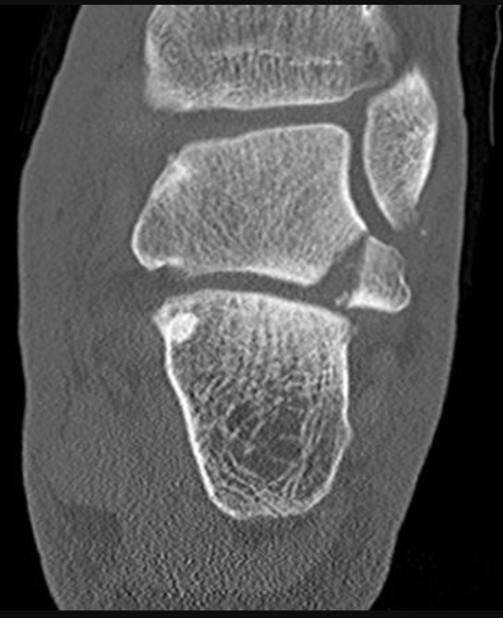
Other injuries occur alongside this fracture in most patients, between about half and nine in ten in published studies. These include cartilage damage in the subtalar joint, loose fragments inside the joint, ligament sprains and dislocation of the peroneal tendons. This is one reason recovery can take longer than expected.1
Types of fracture
- Type I: a single large fragment
- Type II: a fragment broken into several pieces (comminuted)
- Type III: a small chip from the front lower edge2
How far the fragment has moved, and how much of the joint surface it involves, usually matter more for treatment than the type alone.
Treatment
Treatment is tailored to each patient. Most of the evidence comes from small studies, and specialists differ on the exact thresholds, so the details below are a general guide.1,2,7
Treatment without surgery
This is usually suitable for fractures that have not moved (generally less than about 2 mm), small fragments, and chip fractures without other significant joint injuries. It usually involves:
- A cast or boot, with no weight on the foot, for about four to six weeks
- A gradual return to walking
- Repeat imaging to make sure the fragment hasn’t moved
Surgery
Surgery is generally advised for:
- Larger fragments that have moved
- Fractures in several pieces, where small fragments may need removing
- Fractures with related injuries, such as loose fragments, cartilage damage or unstable peroneal tendons
The fragment can be fixed back in place with small screws, or removed if it is too small or too broken to fix. In some cases this can be done by keyhole surgery (ankle arthroscopy).
Displaced fractures appear to do better with early surgery. In one study, two-thirds of patients first treated without surgery went on to need further treatment, and they were more likely to eventually need a fusion of the subtalar joint.7
Recovery after surgery
Timelines vary with the injury and the operation. A typical course is:
- Weeks 0 to 2: a half plaster, no weight on the foot, and keeping the leg raised. Stitches are usually removed at around two weeks.
- Weeks 2 to 6: a cast or boot, still with no weight on the foot.
- Weeks 6 to 10: physiotherapy starts, and weight-bearing is built up step by step in a boot.
- Weeks 12 to 16: back into normal shoes and daily activities, depending on how well the fracture has healed.
- From four to six months: return to running, sports with sudden changes of direction, and snowboarding.
Rehabilitation priorities for physiotherapists
- Restore ankle and especially subtalar movement (inversion and eversion). Subtalar stiffness is common after this injury.
- Control swelling.
- Retrain normal walking once weight-bearing is allowed.
- Strengthen the calf, peroneal and intrinsic foot muscles.
- Retrain balance and joint position sense, progressing to uneven surfaces.
- Add sport-specific training, such as landing and edge control for snowboarders.
Contact the surgeon if there is joint pain that keeps getting worse, catching or locking, new loss of subtalar movement, snapping or clunking of the peroneal tendons, or if progress stalls.
Outlook and long-term problems
Most people do well, especially when the fracture is diagnosed early and treated appropriately. Possible long-term problems include:
- Arthritis in the subtalar joint. This is the most important long-term risk, and the main reason some people have a less good result. In one surgical series, about half of patients developed painful subtalar arthritis.1
- The bone not healing, or healing in the wrong position. This is more likely when the fracture is missed.
- Ongoing pain or pinching on the outside of the ankle, which may need the fragment removing or keyhole surgery.6
- A slower return to sport. In one series, about half of patients returned to their previous level.1
If arthritis pain can’t be managed with simpler measures, a fusion of the subtalar joint may eventually be considered.
Another injury often mistaken for an ordinary sprain is a high ankle sprain (syndesmosis injury).
Referrals and appointments
If you have an ankle injury that isn’t settling, or you are a physiotherapist with a patient you are concerned about, I can see them promptly in London. Request an appointment or message us on WhatsApp.
References
- Hörterer H, Baumbach SF, Lemperle S, et al. Clinical outcome and concomitant injuries in operatively treated fractures of the lateral process of the talus. BMC Musculoskeletal Disorders. 2019.
- Ahmad J, Raikin SM. Current concepts review: talar fractures. Foot & Ankle International. 2006.
- Jentzsch T, Hasler A, Renner N, et al. The V sign in lateral talar process fractures: an experimental study using a foot and ankle model. BMC Musculoskeletal Disorders. 2017.
- Funk JR, Srinivasan SC, Crandall JR. Snowboarder’s talus fractures experimentally produced by eversion and dorsiflexion. American Journal of Sports Medicine. 2003.
- Melenevsky Y, Mackey RA, Abrahams RB, Thomson NB. Talar fractures and dislocations: a radiologist’s guide to timely diagnosis and classification. RadioGraphics. 2015.
- Mui T, et al. Accessory anterolateral talar facet syndrome after treatment of neglected fracture of the lateral process of the talus: a case report. Journal of Foot and Ankle Surgery. 2020.
- Ross H, Marchand L, Cardon J, et al. Patient-reported outcomes after lateral process talus fracture. Journal of Orthopaedic Trauma. 2021.
- Valderrabano V, Perren T, Ryf C, Rillmann P, Hintermann B. Snowboarder’s talus fracture. American Journal of Sports Medicine. 2005.


